Figure 1. [The normal human retina fundus]. - Webvision - NCBI
Por um escritor misterioso
Descrição
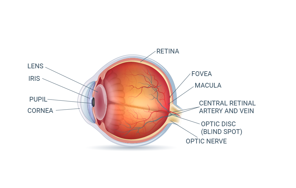
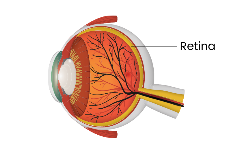
The normal human retina fundus photo shows the optic nerve (right), blood vessels and the position of the fovea (center).
![Figure 1. [The normal human retina fundus]. - Webvision - NCBI](http://webvision.med.utah.edu/wp-content/uploads/2018/05/sagschem.jpg)
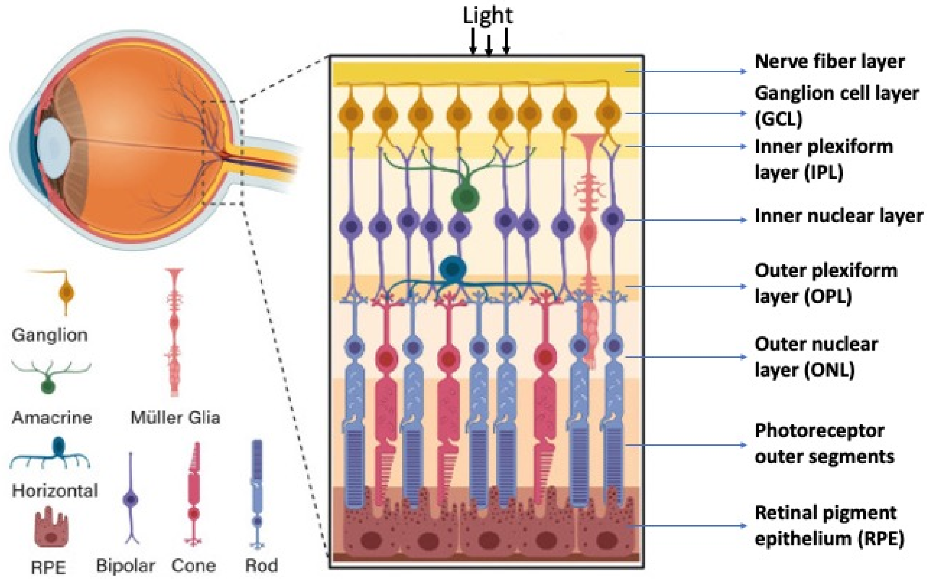
Simple Anatomy of the Retina by Helga Kolb – Webvision
![Figure 1. [The normal human retina fundus]. - Webvision - NCBI](https://eophtha.com/images/uploads/15974738732113548205f378451d43dc.jpg)
Anatomy of Retina
![Figure 1. [The normal human retina fundus]. - Webvision - NCBI](https://media.springernature.com/lw685/springer-static/image/art%3A10.1038%2Fs41467-019-12917-9/MediaObjects/41467_2019_12917_Fig4_HTML.png)
Single-nuclei RNA-seq on human retinal tissue provides improved transcriptome profiling
![Figure 1. [The normal human retina fundus]. - Webvision - NCBI](https://www.biorxiv.org/content/biorxiv/early/2022/02/24/2022.02.22.481546/F1.large.jpg)
Myopia alters the structural organization of the retinal astrocyte template, associated vasculature and ganglion layer thickness
![Figure 1. [The normal human retina fundus]. - Webvision - NCBI](http://webvision.org.es/wp-content/uploads/2017/01/Fig01.png)
Retinal Degeneration, Remodeling and Plasticity. Bryan William Jones, Robert E. Marc and Rebecca L. Pfeiffer - Webvision
![Figure 1. [The normal human retina fundus]. - Webvision - NCBI](https://www.pnas.org/cms/10.1073/pnas.2307380120/asset/116b59e8-9fcc-4213-b822-ca1220677db6/assets/images/large/pnas.2307380120fig02.jpg)
Cellular migration into a subretinal honeycomb-shaped prosthesis for high-resolution prosthetic vision
![Figure 1. [The normal human retina fundus]. - Webvision - NCBI](http://eyerounds.org/atlas/LARGE/Normal-fundus-LRG.jpg)
Atlas Entry - Situs Inversus of the Retinal Vessels
![Figure 1. [The normal human retina fundus]. - Webvision - NCBI](https://webvision.med.utah.edu/wp-content/uploads/2019/07/KrizajFigure4sure.jpg)
What is glaucoma? by David Krizaj – Webvision
![Figure 1. [The normal human retina fundus]. - Webvision - NCBI](https://www.ncbi.nlm.nih.gov/books/NBK11533/bin/muller.gif)
Simple Anatomy of the Retina - Webvision - NCBI Bookshelf
![Figure 1. [The normal human retina fundus]. - Webvision - NCBI](https://www.ncbi.nlm.nih.gov/books/NBK11556/bin/factsf2a.gif)
Facts and Figures Concerning the Human Retina - Webvision - NCBI Bookshelf
![Figure 1. [The normal human retina fundus]. - Webvision - NCBI](https://media.springernature.com/lw685/springer-static/image/chp%3A10.1007%2F978-3-030-25886-3_22/MediaObjects/436773_1_En_22_Fig1_HTML.png)
Image Analysis for Ophthalmology: Segmentation and Quantification of Retinal Vascular Systems
de
por adulto (o preço varia de acordo com o tamanho do grupo)


