Figure 3 from Stimulated Emission Depletion (STED) Microscopy: from Theory to Practice
Por um escritor misterioso
Descrição
Figure 3. Fluorescence depletion of two common dyes in STED microscopy, Atto647N (black, diamonds) and Atto655 (red, circles), as a function of the depletion laser intensity. Error bars for Atto647N appear smaller than the point size of the average value. - "Stimulated Emission Depletion (STED) Microscopy: from Theory to Practice"
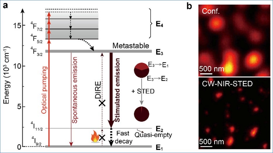
Lanthanide nanoparticles enable continuous-wave NIR STED microscopy

Recent research on stimulated emission depletion microscopy for
Biosensors, Free Full-Text
Super-Resolution Optical Microscopy – Ultrafast and

Ultralow power demand in fluorescence nanoscopy with digitally
ZEISS Microscopy Online Campus Introduction to Superresolution

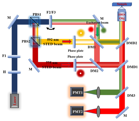
Building a fast scanning stimulated emission depletion microscope

Imaging Living Synapses at the Nanoscale by STED Microscopy
STED Microscopy Scientific Volume Imaging

Fluorescence microscopy with diffraction resolution barrier broken

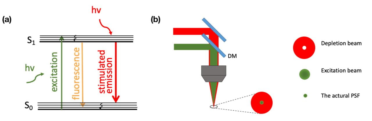
Stimulated Emission Depletion Microscopy

Stimulated Emission Depletion Microscopy
de
por adulto (o preço varia de acordo com o tamanho do grupo)